
Transrectal Ultrasound guided prostate biopsy (TRUS) is an investigation used to diagnose prostate cancer. In general, a TRUS prostate biopsy is performed in men with an abnormal PSA, a palpable abnormality on a digital rectal examination (DRE) or a rapidly increasing PSAlevel.
Transrectal ultrasonography (TRUS)-guided prostate biopsy is the most commonly used method of sampling prostate tissue for the diagnosis of prostate cancer. The technique is well recognised to potentially cause severe pain and discomfort for patients and this has led to numerous attempts to devise ways to minimise these problems. This systematic review summarises the techniques that have been described to date, with special reference to studies using either a visual analogue or numerical analogue scale to report outcomes. Commonly used approaches that are effective to minimise pain or discomfort include intravenous sedoanalgesia, inhalational agents and periprostatic infiltration of local anaesthetic. Whilst diclofenac suppositories are more effective than placebo, intra-rectal local anaesthetic gels appear to be of no benefit. Performing TRUS-guided prostate biopsy without any form analgesia is not appropriate
he doctor or nurse uses a thin needle to take small samples of tissue from the prostate.
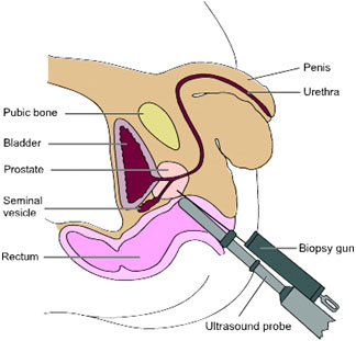
You’ll lie on your side on an examination table, with your knees brought up towards your chest. The doctor or nurse will put an ultrasound probe into your back passage (rectum), using a gel to make it more comfortable. The ultrasound probe scans the prostate and an image appears on a screen. The doctor or nurse uses this image to guide where they take the cells from. If you’ve had an MRI scan, the doctor or nurse may use the images to decide which areas of the prostate to take biopsy samples from.
You will have an injection of local anaesthetic to numb the area around your prostate and reduce any discomfort. The doctor or nurse then puts a needle next to the probe in your back passage and inserts it through the wall of the back passage into the prostate. They usually take 10 to 12 small pieces of tissue from different areas of the prostate. But, if the doctor is using the images from your MRI scan to guide the needle, they may take fewer samples.